Powder: -20°C for 3 years | In solvent: -80°C for 1 year
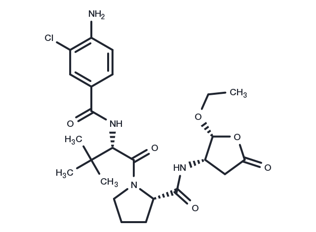
Belnacasan (VX-765) 是一种具有口服活性的 IL 转换酶/caspase-1 抑制剂,是 VRT-043198 的口服生物活性前药,作用于外周血单核细胞,可抑制 LPS 诱导的 IL-1β 和 IL-18 释放。

| 规格 | 价格/CNY | 货期 | 数量 | |
|---|---|---|---|---|
| 1 mg | ¥ 276 | 现货 | ||
| 2 mg | ¥ 393 | 现货 | ||
| 5 mg | ¥ 647 | 现货 | ||
| 10 mg | ¥ 898 | 现货 | ||
| 25 mg | ¥ 1,780 | 现货 | ||
| 50 mg | ¥ 2,990 | 现货 | ||
| 100 mg | ¥ 3,850 | 现货 | ||
| 200 mg | ¥ 5,520 | 现货 | ||
| 1 mL * 10 mM (in DMSO) | ¥ 762 | 现货 | ||
| 产品描述 | Belnacasan (VX-765) is an orally active IL-converting enzyme/caspase-1 inhibitor. |
| 靶点活性 | Caspase-1:0.8 nM(Ki), Caspase-4:<0.6 nM(Ki) |
| 体外活性 | VX-765通过防止前脑星形胶质细胞中IL-1β的增加阻断大鼠体内的癫痫发生,而对后放电持续时间没有明显影响.在患有遗传性失神癫痫的成年大鼠体内注射3天VX-765后,通过选择性阻断IL-1β生物合成,明显降低累积持续时间,减少平均55%的棘慢波放电.50 mg/kg-200 mg/kg VX-765在急性癫痫小鼠模型中,通过延迟首次癫痫开始时间,并减少平均50%的癫痫发作次数以及64%的总持续时间,产生抗癫痫作用.200 mg/kg VX-765在胶原诱导的关节炎小鼠模型中,抑制60%脂多糖诱导的IL-1β产生,并导致炎症评分剂量依赖性明显降低,有效保护关节病变. |
| 体内活性 | VRT-043198会抑制IL-1β从外周血单个核细胞和全血中的释放,IC50分别为0.67 μM和1.9 μM。VX-765是一种口服可吸收的VRT-043198前药,显示出对ICE /半胱天冬酶-1和半胱天冬酶-4的有效抑制作用,Ki分别为0.8 nM和<0.6 nM。 |
| 激酶实验 | Enzyme inhibition is assayed by tracking of the rate of hydrolysis of an appropriate substrate labeled with either p-nitroaniline or aminomethyl coumarin (AMC) as follows: ICE/caspase-1, suc-YVAD-p-nitroanilide; caspase-4, Ac-WEHD-AMC; caspase-6, Ac-VEID-AMC; caspase-3, -7, -8, and -9, Ac-DEVD-AMC; and granzyme B, Ac-IEPD-AMC. Enzymes and substrates are incubated in a reaction buffer [10 mM Tris, pH 7.5, 0.1% (w/v) CHAPS, 1 mM dithiothreitol, and 5% (v/v) DMSO] for 10 min at 37°C. Glycerol is added to the buffer at 8% (v/v) for caspase-3, -6, and -9 and granzyme B to improve stability of enzymes. The rate of substrate hydrolysis is monitored using a fluorometer. Assays for cathepsin B and trypsin are performed[2]. |
| 细胞实验 | VX-765 is solubilized in DMSO and stored, and then diluted with RPMI 1640 complete medium (DMSO 0.2%) before use[1]. A total of 2×10⁵ cells/well (100 μL cell suspension) is distributed in triplicate in flat-bottom 96-well plates. Either 50 μL of VX-765 (40 μM in RPMI 1640 complete medium containing 0.2% DMSO) or vehicle control is added to appropriate wells. Following a 30-min incubation at 37°C, 50 μL of LPS diluted in RPMI 1640 complete medium is added at final concentrations varying from 0.001 to 10 ng/mL. Cells are returned to a 37°C incubator. At 4 h after LPS addition, 75 μL of supernatant is removed from wells, cleared by centrifugation for 5 min at 1500 rpm, and stored at 4°C until assayed. Cells are returned to a 37°C incubator until 24 h after LPS addition, at which time 100 μL of supernatant is removed, cleared by centrifugation, and stored at 4°C. Supernatants are tested using ELISA kits for IL-1β, IL-6, IL-18, and IL-1α[1]. |
| 别名 | VX-765 |
| 分子量 | 508.99 |
| 分子式 | C24H33ClN4O6 |
| CAS No. | 273404-37-8 |
Powder: -20°C for 3 years | In solvent: -80°C for 1 year
Ethanol: 93 mg/mL (182.7 mM)
DMSO: 93 mg/mL (182.7 mM)
| 可选溶剂 | 浓度 体积 质量 | 1 mg | 5 mg | 10 mg | 25 mg |
| Ethanol / DMSO | 1 mM | 1.9647 mL | 9.8234 mL | 19.6468 mL | 49.1169 mL |
| 5 mM | 0.3929 mL | 1.9647 mL | 3.9294 mL | 9.8234 mL | |
| 10 mM | 0.1965 mL | 0.9823 mL | 1.9647 mL | 4.9117 mL | |
| 20 mM | 0.0982 mL | 0.4912 mL | 0.9823 mL | 2.4558 mL | |
| 50 mM | 0.0393 mL | 0.1965 mL | 0.3929 mL | 0.9823 mL | |
| 100 mM | 0.0196 mL | 0.0982 mL | 0.1965 mL | 0.4912 mL |
对于不同动物的给药剂量换算,您也可以参考 更多...
请在以下方框中输入您的动物实验信息后点击计算,可以得到母液配置方法和体内配方的制备方法: 比如您的给药剂量是10 mg/kg,每只动物体重20 g,给药体积100 μL,一共给药动物10 只,您使用的配方为5% DMSO+30% PEG300+5% Tween 80+60% ddH2O。那么您的工作液浓度为2 mg/mL。
母液配置方法:2 mg 药物溶于 50 μL DMSO (母液浓度为 40 mg/mL), 如您需要配置的浓度超过该产品的溶解度,请先与我们联系。
体内配方的制备方法:取 50 μL DMSO 主液,加入 300 μL PEG300, 混匀澄清,再加 50 μL Tween 80,混匀澄清,再加 600 μL ddH2O, 混匀澄清。
您可能有的问题的答案可以在抑制剂处理说明中找到,包括如何准备库存溶液,如何存储产品,以及基于细胞的分析和动物实验需要特别注意的问题。
Belnacasan 273404-37-8 Apoptosis Proteases/Proteasome Caspase Inhibitor VX 765 VX765 inhibit VX-765 inhibitor
